
Following worms are described below:
Liver flukes, Tape worms, Lung worms, Round worms
For Eye worms - see under Eye problems
Liver Flukes
Local names:
Embu: nthambara / Gikuyu: thambara cia mani / Kamba: ntambaa / Kipsigis: sungurutek / Luo: ochwe / Maragoli: ovoveyi / Meru: nthanthara / Samburu: ikurui, lemonyua / Somali Ethiopia: faraqle / Somali Kenya: sogul / Masai: Osinkirri
Family: Trematoda
Description: Parasite
Introduction
There are two important species of liver flukes in Kenya: fasciola gigantica and fasciola hepatica. The former is more important, being found throughout the lower warmer parts of the country. As the name suggests it is very large, more than twice the size of fasciola hepatica, which is found in the cooler highland areas of the country, and also in the temperate zones of the world such as Europe, North America, Australia and New Zealand.
The life cycle of liver flukes involves a snail, which acts as an intermediate host. If there is no snail there will be no flukes. The snail involved requires still, stagnant water to survive; swift running water does not suit it. Swamps, ponds, lakes with a marshy edge and pools of water edged by vegetation are danger zones for grazing animals.
Cattle, sheep and goats are mainly affected, although other animals such as donkeys, horses, and many species of wildlife such as buffalo can also be infested. Humans can also be infested with liver flukes.
As the name suggests, the liver fluke attacks the liver.
Three forms of illness can occur:
- Flukes can cause sudden death, from liver failure and from internal bleeding, when large numbers of immature flukes migrate through and cause damage to liver tissue. This clinical picture is common in young sheep.
- Flukes can also cause a chronic wasting disease accompanied by anaemia and oedema (swelling). The oedema is typically located on the lower jaws (“bottle jaw”) and on the lower part of the belly. This is the most common picture in cattle.
- Liver lesions due to flukes are the causative factor in Infectious Necrotic Hepatitis (Black Disease), predominantly a disease of sheep aged between 2 - 4 years, but aso occurs in young cattle. Specific bacteria (Clostridia) multiply in liver lesions caused by migrating flukes and release toxins. Sudden death is the result.
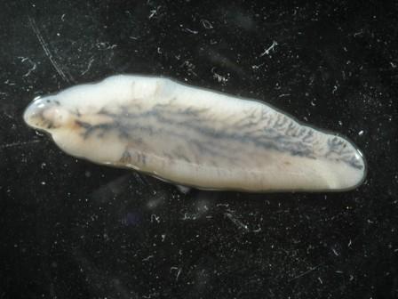 |
| Adult of Fasciola hepatica |
|
© Wikipedia
|
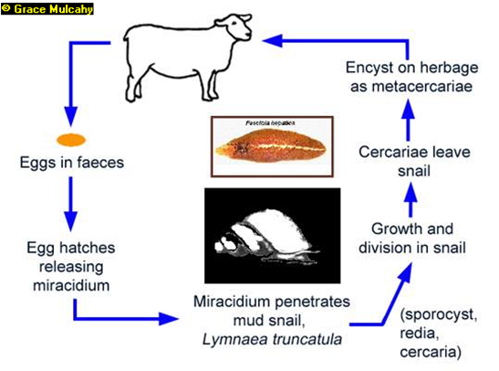
Life cycle of liver flukes
Knowledge of the life cycle of the liver fluke is an aid in understanding how to control the disease.
The intermediate host snails for Fasciola gigantica and Fasciola hepatica may differ but in other respects the life cycle is the same.
- The life cycle begins with the eggs of the fluke which mature in the bile ducts in the liver, pass down the ducts, into the gut and are excreted with the faeces.
- Once outside in the environment, which must contain water, the eggs hatch, releasing an active stage, called miracidia. Temperature and time are critical in the early stages for the development of the miracidia- above 5-6 C, and best between 25-24C. Miracidia must find a suitable snail within 24-30 hours or they will die.
- The miracidia either actively invade a host snail or are eaten by a host snail.
- They then hatch in the snail’s gut and the next stage develops in the tissues of the snail.
- 5 to 8 weeks later another stage emerges from the snail and form resistant cysts attached to herbage or grass, where they are eaten by the final host – cattle, sheep, goat or other herbivores.
- Once ingested by the sheep or cow the immature fluke invade the gut wall, travel to the liver where they cause extensive damage to the liver tissue until they reach the bile ducts. Here they mature into adult flukes and start to lay eggs and the life cycle begins again. It takes 10-12 weeks from infestation until eggs start to be laid and shed with the faeces.
Mature flukes are long lived and sheep and cattle may be carriers for years.
 |
|
Lifecycle of Fasciola hepatica |
|
© Grace Mulcahy
|
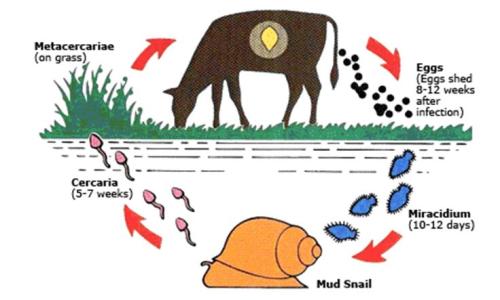 |
| Life cycle of a liver fluke |
|
© uk.merial.com
|
Signs of Liver Fluke disease
The severity of clinical signs depends on the number of parasites ingested by the host animal over a short period of time. Cattle appear to be able to develop an immune reaction to fluke infestation, but sheep do not.
The acute form of the disease is more common in sheep than in cattle.
- It occurs 5 to 6 weeks after the ingestion of large numbers of metacercariae. There is a sudden invasion of the liver by masses of young liver flukes.
- Sudden death, especially in sheep, can occur due to internal bleeding and liver failure and also due to sudden release of toxin by bacteria (Clostridia) multiplying rapidly in the liver lesions (Black Disease)
- The acute form of the disease in cattle is manifested either by sudden death, or dullness, weakness, anaemia, lack of appetite, pain over the region of the liver and death in 48 hours.
- Because this is caused by migrating flukes which have not yet begun to lay eggs, no eggs are found in the faeces at laboratory analysis; diagnosis is only possible by post mortem examination.
- Post mortem will reveal a badly damaged, swollen liver with many small perforations and haemorrhages (internal bleeding spots). The liver tissue contains mini-tunnels formed by the migrating immature flukes, which are visible by the naked eye at careful close examination.
The sub-acute or chronic form of the disease is more common in cattle, but also occurs in sheep and can cause death in both. It is the result of activity of the adult flukes in the bile ducts, causing anaemia and protein leakage.
- Body growth is reduced, there is loss of weight and often emaciation, oedema appear on the lower jaw (`bottle jaw`) and on the lower belly, there can be anaemia.
- Diagnosis in the laboratory is made by finding fluke eggs in faecal samples.
- A post mortem examination will reveal large mature flukes in the bile ducts, which in cattle are usually thickened and may be hardened in cattle, these bile duct changes are not seen in sheep.
Diagnosis
Acute fluke infestation usually kills younger animals, especially sheep. Age difference is helpful in making a diagnosis based on the clinical signs, grazing history, access to stagnant water, season (whether wet or dry), post mortem findings, the finding of immature or mature flukes and the demonstration of fluke eggs in faecal samples.
Prevention – Control - Treatment
Prevention and control
- Control measures include treatment against flukes in affected animals and the prevention of livestock access to snail-infested pastures. In practice only the first of these is used in most cases.
- Fencing off snail sites makes sense when the areas are small. Preventing livestock from drinking from ponds and stagnant water where they can feed from peri-aquatic vegetation infested by liver fluke cysts is an important measure. This an bee achieved by providing adequately maintained water troughs for watering of livestock with a good concreted area around them to prevent the area from becoming muddied.
- Drainage of pastures and paddocks is the best permanent solution, but may be costly.
- Grazing livestock in Africa are more likely to contract liver flukes infestations during the dry season than in the wet, as during the dry season they enter snail infested flood zones and marshes which constitute dry season grazing reserves. In such situations it is advisable to keep animals out of such swamps for as long as possible so that fewer viable cysts are present on the herbage.
- When entry to the swamp is unavoidable then the older cattle should go in first as they are much more resistant than the younger cattle, followed, last of all, by the most susceptible sheep and goats.
- The prophylactic administration of specific drugs against flukes to those animals most at risk, can considerably reduce occurrence of the disease.
Treatment
The following drugs are effective in the treatment of Liver Flukes:
- Triclabendazole and Fenbendazole should be given at the rate of 10mg and 8mg respectively per kg body weight by mouth.
- Trodax or nitroxynil given at 34 % solution for cattle administered subcutenously at 1.5ml per 50kg body weight and may be repeated as may be necessary.
- Oxyclozanide or Flukanide or Ranide are available from different pharmaceutical manufacturers and should be used according to the manufacturer’s recommendation.
Review process:
1. William Ayako, KARI Naivasha. Aug - Dec 2009
2. Hugh Cran , Practicing Veterinarian Nakuru. March - Oct 2010
3. Review workshop team. Nov 2 - 5, 2010
- For Infonet: Anne, Dr Hugh Cran
- For KARI: Dr Mario Younan KARI/KASAL, William Ayako - Animal scientist, KARI Naivasha
- For DVS: Dr Josphat Muema - Dvo Isiolo, Dr Charity Nguyo - Kabete Extension Division, Mr Patrick Muthui - Senior Livestock Health Assistant Isiolo, Ms Emmah Njeri Njoroge - Senior Livestock Health Assistant Machakos
- Pastoralists: Dr Ezra Saitoti Kotonto - Private practitioner, Abdi Gollo H.O.D. Segera Ranch
- Farmers: Benson Chege Kuria and Francis Maina Gilgil and John Mutisya Machakos
- Language and format: Carol Gachiengo
Information Source Links
- Barber, J., Wood, D.J. (1976) Livestock management for East Africa: Edwar Arnold (Publishers) Ltd 25 Hill Street London WIX 8LL
- Blood, D.C., Radostits, O.M. and Henderson, J.A. (1983) Veterinary Medicine - A textbook of the Diseases of Cattle, Sheep, Goats and Horses. Sixth Edition - Bailliere Tindall London. ISBN: 0702012866
- Blowey, R.W. (1986). A Veterinary book for dairy farmers: Farming press limited Wharfedale road, Ipswich, Suffolk IPI 4LG
- Force, B. (1999). Where there is no Vet. CTA, Wageningen, The Netherlands. ISBN 978-0333-58899-4
- Hall, H.T.B. (1985). Diseases and parasites of Livestock in the tropics. Second Edition. Longman Group UK. ISBN 0582775140
- Handbook on Animal Diseases in the Tropics 4th Edition Sewell & Brocklesby
- Hunter, A. (1996). Animal health: General principles. Volume 1 (Tropical Agriculturalist) - Macmillan Education Press. ISBN: 0333612027
- Hunter, A. (1996). Animal health: Specific Diseases. Volume 2 (Tropical Agriculturalist) - Macmillan Education Press. ISBN:0-333-57360-9
- ITDG and IIRR (1996). Ethnoveterinary medicine in Kenya: A field manual of traditional animal health care practices. Intermediate Technology Development Group and International Institute of Rural Reconstruction, Nairobi, Kenya. ISBN 9966-9606-2-7.
- Merck Veterinary Manual 9th Edition
- Pagot, J. (1992). Animal Production in the Tropics and Subtropics. MacMillan Education Limited London
- The Organic Farmer magazine No. 50 July 2009
- The Organic Farmer magazine No. 51 August 2009